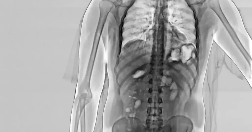
Pastrana X-Ray: Unveiling the Science Behind the Stunts and Injuries
Travis Pastrana, a name synonymous with extreme sports, gravity-defying stunts, and a relentless pursuit of pushing boundaries, has captivated audiences for decades. But behind the roaring engines and spectacular aerial maneuvers lies a less glamorous reality: the inevitable toll on the human body. This is where the Pastrana X-Ray comes into play, offering a glimpse into the internal consequences of a career lived on the edge. This article delves into the science behind the Pastrana X-Ray, exploring its significance in understanding and managing the injuries sustained by extreme athletes like Travis Pastrana.
The Inevitable Impact: Understanding Extreme Sports Injuries
Extreme sports, by their very nature, involve high levels of risk. The combination of speed, height, and complex maneuvers creates a perfect storm for injuries. From motocross and rally racing to base jumping and freestyle skiing, athletes like Pastrana constantly expose themselves to significant forces and potential trauma. These forces can result in a wide range of injuries, including fractures, dislocations, sprains, strains, and concussions. The repetitive nature of training and competition can also lead to chronic conditions like osteoarthritis and tendinitis. The Pastrana X-Ray becomes a crucial tool in diagnosing and monitoring these conditions.
The Role of X-Rays in Diagnosing and Monitoring Injuries
X-rays, also known as radiographs, are a form of electromagnetic radiation that can penetrate soft tissues but are absorbed by denser materials like bone. This allows medical professionals to create images of the skeletal system, revealing fractures, dislocations, and other abnormalities. In the context of extreme sports, Pastrana X-Ray imaging plays a vital role in several key areas:
- Diagnosis of Acute Injuries: When an athlete sustains a sudden injury, such as a fall or collision, an X-ray can quickly identify fractures, dislocations, and other structural damage. This information is essential for determining the appropriate course of treatment.
- Monitoring Healing Progress: After an injury has been treated, X-rays can be used to monitor the healing process. This allows doctors to assess whether a fracture is healing properly, whether a joint is stable, and whether there are any signs of complications.
- Identifying Chronic Conditions: Repetitive stress and trauma can lead to chronic conditions like osteoarthritis and tendinitis. X-rays can help identify these conditions by revealing changes in bone structure, joint space narrowing, and other signs of wear and tear.
- Pre-Participation Screening: Some athletes undergo pre-participation X-rays to identify any pre-existing conditions that could increase their risk of injury. This information can be used to develop individualized training programs and injury prevention strategies.
Beyond the Basics: Advanced Imaging Techniques
While X-rays are a valuable diagnostic tool, they have limitations. They are best suited for visualizing bone structures and may not provide detailed information about soft tissues like ligaments, tendons, and muscles. In some cases, more advanced imaging techniques may be necessary to fully evaluate an injury. These techniques include:
- Magnetic Resonance Imaging (MRI): MRI uses magnetic fields and radio waves to create detailed images of soft tissues. It is particularly useful for diagnosing ligament tears, tendon ruptures, and other soft tissue injuries.
- Computed Tomography (CT) Scans: CT scans use X-rays to create cross-sectional images of the body. They provide more detailed information about bone structures than traditional X-rays and can also be used to visualize soft tissues.
- Ultrasound: Ultrasound uses sound waves to create images of soft tissues. It is a non-invasive and relatively inexpensive imaging technique that is often used to evaluate tendon and muscle injuries.
The choice of imaging technique depends on the specific injury being evaluated and the information needed to make an accurate diagnosis. In many cases, a combination of imaging techniques may be used to provide a comprehensive assessment. When it comes to a prominent figure like Travis Pastrana, access to these advanced imaging technologies is paramount for his continued performance and safety.
Travis Pastrana: A Case Study in Extreme Sports Injuries
Travis Pastrana’s career has been marked by a series of spectacular successes and equally dramatic injuries. He has broken numerous bones, suffered multiple concussions, and undergone countless surgeries. His willingness to push the limits has made him a legend in the world of extreme sports, but it has also taken a significant toll on his body. A hypothetical Pastrana X-Ray collection over his career would be a fascinating, if sobering, study of the impact of extreme sports on the musculoskeletal system. The insights gained from analyzing his injuries could inform injury prevention strategies for other athletes.
Pastrana’s injuries are a testament to the forces involved in his chosen disciplines. From high-speed crashes on motorcycles to bone-jarring landings in rally cars, his body has been subjected to immense stress. The Pastrana X-Ray images would likely reveal a history of fractures, dislocations, and other injuries in various parts of his body. They might also show signs of chronic conditions like osteoarthritis, which can develop as a result of repetitive trauma.
Understanding the nature and extent of Pastrana’s injuries is crucial for developing effective treatment and rehabilitation strategies. It can also help him make informed decisions about his future participation in extreme sports. While he has shown an incredible ability to recover from injuries, it is important to recognize that there are limits to what the human body can withstand. The information gleaned from Pastrana X-Ray examinations, combined with other diagnostic tools, provides a comprehensive picture of his physical condition.
Injury Prevention Strategies in Extreme Sports
While injuries are an inherent risk in extreme sports, there are steps that athletes can take to minimize their risk. These strategies include:
- Proper Training and Conditioning: Athletes should undergo rigorous training programs to strengthen their muscles, improve their flexibility, and enhance their coordination. This can help them better withstand the forces involved in extreme sports.
- Using Appropriate Protective Gear: Protective gear, such as helmets, pads, and braces, can help absorb impact and protect against injuries. It is essential to use high-quality protective gear that is specifically designed for the sport being practiced.
- Avoiding Overexertion: Athletes should avoid pushing themselves too hard, especially when they are tired or injured. Overtraining can increase the risk of injury and slow down the healing process.
- Listening to Their Bodies: Athletes should pay attention to their bodies and recognize the signs of pain and fatigue. Ignoring these signals can lead to more serious injuries.
- Regular Medical Checkups: Regular medical checkups can help identify potential problems before they become serious. Athletes should consult with a sports medicine physician or other qualified healthcare professional to develop an individualized injury prevention plan.
The Future of Injury Management in Extreme Sports
The field of sports medicine is constantly evolving, with new technologies and treatments being developed all the time. In the future, we can expect to see even more sophisticated methods for diagnosing, treating, and preventing injuries in extreme sports. These advancements will include:
- Improved Imaging Techniques: New imaging techniques, such as 3D X-rays and advanced MRI sequences, will provide even more detailed information about injuries. This will allow doctors to make more accurate diagnoses and develop more targeted treatment plans.
- Regenerative Medicine: Regenerative medicine therapies, such as stem cell injections and platelet-rich plasma (PRP) therapy, have the potential to accelerate the healing process and promote tissue regeneration. These therapies are showing promise for treating a variety of sports-related injuries.
- Personalized Medicine: Personalized medicine approaches will take into account an individual’s genetic makeup, lifestyle, and other factors to develop customized treatment and prevention plans. This will allow athletes to receive the most effective and appropriate care possible.
The Pastrana X-Ray and the data it provides, combined with advances in sports medicine, will play a crucial role in ensuring the safety and longevity of athletes in extreme sports. By understanding the risks involved and implementing effective injury prevention strategies, we can help these athletes continue to push the limits of human performance while minimizing the potential for long-term health consequences. [See also: Concussion Protocols in Extreme Sports] [See also: The Role of Physical Therapy in Injury Recovery]
Conclusion: The Enduring Legacy of Extreme Sports and the Importance of Medical Advancements
Extreme sports continue to captivate audiences worldwide, showcasing the incredible athleticism and daring spirit of individuals like Travis Pastrana. However, the inherent risks associated with these activities necessitate a comprehensive understanding of potential injuries and effective management strategies. The Pastrana X-Ray, as a representation of diagnostic imaging, serves as a critical tool in this process, allowing medical professionals to assess the extent of injuries and guide treatment plans. As sports medicine continues to advance, with improved imaging techniques and personalized approaches to care, the future looks promising for athletes seeking to push their boundaries while minimizing the long-term impact on their bodies. The legacy of extreme sports will be shaped not only by the spectacular feats of athleticism but also by the dedication to athlete safety and well-being.
