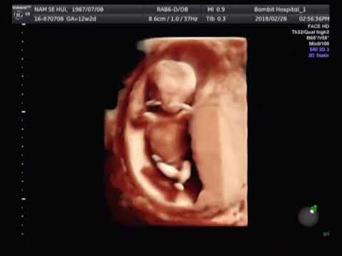
Decoding the 12 Week Old Baby Ultrasound: A Comprehensive Guide
The 12 week old baby ultrasound is a pivotal moment in prenatal care, offering expectant parents a first glimpse of their developing child. This ultrasound, typically performed between 11 and 14 weeks of gestation, provides crucial information about the baby’s health, development, and estimated due date. Understanding what to expect during a 12 week old baby ultrasound and how to interpret the results can alleviate anxiety and foster a stronger connection with the growing baby.
What is a 12 Week Old Baby Ultrasound?
A 12 week old baby ultrasound, also known as a nuchal translucency (NT) scan, is a non-invasive imaging technique that uses high-frequency sound waves to create real-time images of the fetus. This ultrasound is generally performed transabdominally, where a transducer is moved across the mother’s abdomen. In some cases, a transvaginal ultrasound may be necessary for a clearer image, especially if the mother has a tilted uterus or if the baby is positioned in a way that makes visualization difficult.
Purpose of the 12 Week Ultrasound
The primary purposes of a 12 week old baby ultrasound include:
- Confirming Pregnancy and Gestational Age: The ultrasound confirms the pregnancy and accurately determines the gestational age of the fetus, which is crucial for calculating the estimated due date.
- Assessing Fetal Viability: The presence of a heartbeat confirms that the fetus is alive.
- Detecting Multiple Pregnancies: The ultrasound can identify if the mother is carrying twins, triplets, or more.
- Evaluating Fetal Anatomy: The sonographer will examine the baby’s basic anatomy, including the head, limbs, and abdomen, to look for any major abnormalities.
- Measuring Nuchal Translucency: This measurement is a key component of the first trimester screening for chromosomal abnormalities such as Down syndrome (Trisomy 21), Trisomy 18 (Edwards syndrome), and Trisomy 13 (Patau syndrome).
What to Expect During the Ultrasound Procedure
Before the 12 week old baby ultrasound, you may be asked to drink water to fill your bladder, which helps improve the image quality. During the procedure, you will lie on your back on an examination table. A gel will be applied to your abdomen to help the transducer glide smoothly and improve sound wave transmission. The sonographer will move the transducer across your abdomen, capturing images of the fetus. The procedure typically takes about 20-30 minutes.
You may be able to see the baby on the screen, and the sonographer may point out various anatomical features. It’s important to remember that the sonographer is primarily focused on obtaining accurate measurements and assessing the baby’s health. They may not be able to provide detailed explanations of everything you see.
Understanding the Ultrasound Report
The ultrasound report will include various measurements and observations. Key components include:
- Crown-Rump Length (CRL): This measurement is the length of the fetus from the top of the head (crown) to the bottom of the buttocks (rump). It is used to accurately determine gestational age.
- Fetal Heart Rate (FHR): This measures the number of times the baby’s heart beats per minute. A normal fetal heart rate at 12 weeks is typically between 120 and 160 beats per minute.
- Nuchal Translucency (NT): This is the measurement of the fluid-filled space at the back of the baby’s neck. An increased NT measurement can indicate an increased risk of chromosomal abnormalities.
- Nasal Bone: The presence or absence of the nasal bone is also assessed, as its absence can be associated with certain chromosomal conditions.
- Basic Anatomy: The sonographer will note the appearance of the baby’s head, brain, limbs, and other major organs.
Interpreting Nuchal Translucency (NT) Measurements
The NT measurement is a critical component of the 12 week old baby ultrasound. The NT measurement is considered increased if it is above a certain threshold, typically 3.5 mm. However, the normal range can vary slightly depending on the gestational age. An increased NT measurement does not necessarily mean that the baby has a chromosomal abnormality. It simply indicates an increased risk, and further testing, such as chorionic villus sampling (CVS) or amniocentesis, may be recommended to confirm the diagnosis. [See also: Understanding Chorionic Villus Sampling]
It is important to discuss the NT measurement and the associated risks with your healthcare provider. They can provide personalized guidance based on your individual circumstances and medical history.
What if Abnormalities are Detected?
If the 12 week old baby ultrasound reveals any abnormalities, it is natural to feel anxious and concerned. However, it is important to remember that many abnormalities detected during ultrasound are not serious and may resolve on their own. In other cases, further testing and monitoring may be necessary to determine the severity of the condition and to develop a plan of care.
Your healthcare provider will discuss the findings with you in detail and explain the available options. These may include:
- Further Ultrasound Scans: Serial ultrasounds may be performed to monitor the baby’s growth and development.
- Genetic Counseling: A genetic counselor can provide information about the risks of chromosomal abnormalities and discuss the available testing options.
- Chorionic Villus Sampling (CVS): This is a diagnostic test that involves taking a small sample of the placenta to analyze the baby’s chromosomes.
- Amniocentesis: This is another diagnostic test that involves taking a sample of the amniotic fluid to analyze the baby’s chromosomes.
- Fetal Echocardiogram: This is a specialized ultrasound that examines the baby’s heart in detail.
The Emotional Impact of the 12 Week Ultrasound
The 12 week old baby ultrasound is a significant milestone in pregnancy. For many parents, it is the first time they see their baby and hear the heartbeat. This can be an emotional and bonding experience. However, it is also a time of potential anxiety and uncertainty. It is important to allow yourself to feel your emotions and to seek support from your partner, family, and healthcare provider. [See also: Coping with Pregnancy Anxiety]
Remember that the vast majority of 12 week old baby ultrasounds are normal, and the baby is healthy and developing as expected. Even if abnormalities are detected, there are often options for treatment and management. Staying informed, communicating openly with your healthcare provider, and seeking emotional support can help you navigate this journey with confidence and peace of mind.
Benefits of Early Ultrasound Screening
Undergoing a 12 week old baby ultrasound offers numerous benefits, including:
- Early Detection of Potential Issues: Identifying potential problems early allows for timely intervention and management, improving outcomes for both mother and baby.
- Accurate Dating of Pregnancy: Knowing the accurate gestational age is crucial for monitoring fetal growth and development.
- Reduced Anxiety: Seeing the baby and hearing the heartbeat can reassure parents and reduce anxiety about the pregnancy.
- Informed Decision-Making: Having information about the baby’s health allows parents to make informed decisions about their prenatal care and delivery options.
- Strengthened Bonding: The ultrasound experience can foster a stronger connection between parents and their baby.
Conclusion
The 12 week old baby ultrasound is an essential part of prenatal care, providing valuable information about the baby’s health and development. Understanding what to expect during the procedure and how to interpret the results can empower expectant parents to make informed decisions and to feel more connected to their growing child. While the experience can be emotional, remember that the vast majority of ultrasounds are normal, and any concerns can be addressed with the support of your healthcare provider. A healthy 12 week old baby ultrasound is a great step towards a healthy pregnancy.
